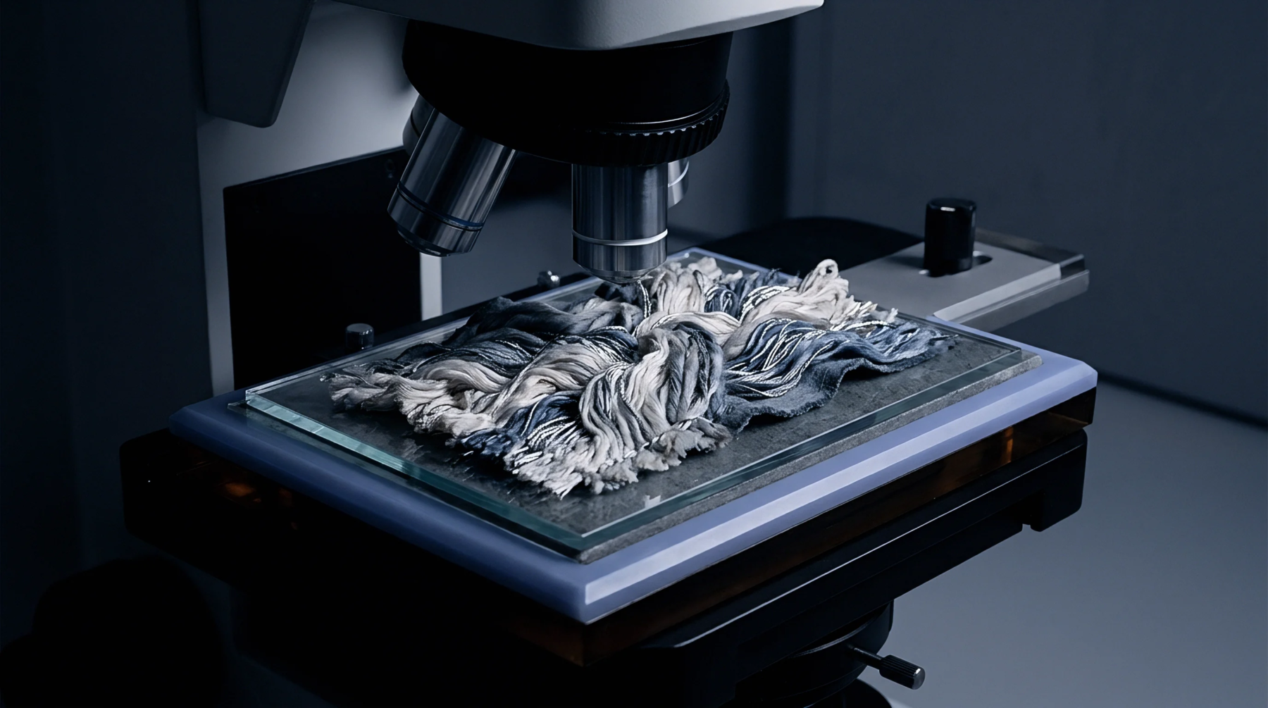
Analytical Modality
Magnification instrumentation utilizing visible light and glass optical lenses constitutes the standard reference platform for textile fibre identification and dimensional analysis. Transmitted illumination passes through prepared specimens, forming magnified images through objective and eyepiece lens combinations at magnifications from 100x to 1000x. The technique identifies longitudinal surface structures, internal pigment distribution, and cross-sectional geometries across animal, vegetable, and synthetic fibres.
Light microscopy establishes the benchmark measurements cited in trade rules for fiber fineness, blend quantification, and species verification. The method stops delivering useful diagnostic information when dealing with sub-micron surface details, structural features below the diffraction limit of light, or heavily dyed opaque fibres that block light transmission entirely.
Measurement Protocol
Standard test methods designate projection light microscopes as the primary apparatus for measuring mean fibre diameter and scale metrics. An operator mounts cut fiber snippets in a refractive liquid beneath a glass cover slide on a movable mechanical stage. The instrument projects the magnified silhouette onto a calibrated circular screen or digital charge-coupled device sensor displaying a measurement grid.
The technician measures the width of each focused snippet across its central axis, recording hundreds of data points to generate statistical distribution curves. Magnification must be calibrated precisely against a certified stage micrometer before each testing session, as minor mechanical shifts in optical tube length produce systematic errors across large sample sets.
Diagnostic Distinctions
Transmitted illumination reveals internal structural features that distinguish natural fibres from manufactured synthetics. Cotton displays collapsed, twisted ribbon structures with visible lumina, whereas flax presents cross-markings known as nodes or dislocations along cylindrical cell walls. Animal fibres show clear cuticular scales along their outer perimeters, occasionally accompanied by continuous, interrupted, or fragmented central medullae.
Cashmere reveals thin, faint scale margins with low profile heights, whereas fine sheep wool presents distinct, serrated scale bands under identical light conditions. Cross-sectional examinations accomplished through wedge-cutting plates further differentiate round fibres, serrated viscose cross-sections, and trilobal synthetic profiles, resolving processing and identity questions that flat longitudinal views cannot settle.
Commercial Significance
Inspection reports generated through light microscope evaluations govern international trade disputes regarding adulterated textile lots. Commercial purchase contracts require mill deliveries to match documented fiber blend declarations within specified regulatory tolerances. When an importer suspects that a cashmere shipment contains cheaper fine sheep wool, light microscope scale counts provide the initial legal evidence for commercial claims.
Automated optical analyzers have accelerated high-volume mill throughput, but manual optical microscopy retains its status as the legal reference arbiter when dispute clauses trigger re-testing. Mill claims regarding fabric composition fail compliance checks if optical cross-section assessments show prohibited synthetic or coarse animal fractions within the spun yarn.