Enzymatic Tryptic Digestion Kinetic Variance in Severely Damaged Animal Fiber Matrices
Enzymatic tryptic digestion of damaged animal fibers requires kinetic rate normalization to prevent severe quantitative misclassification in fiber identification

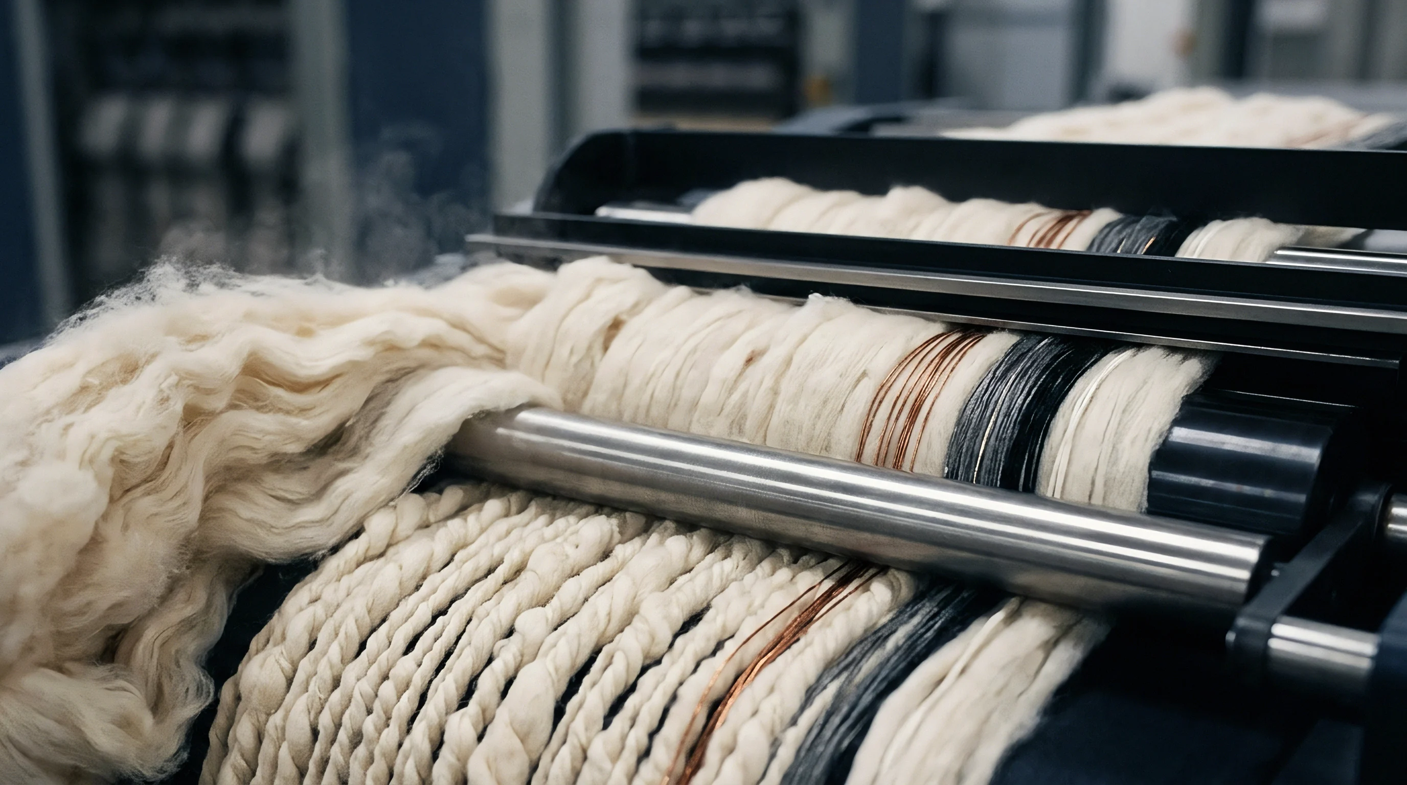
Cleave
In animal keratin fibers, proteins form a dense, three-dimensional network crosslinked by disulfide bridges, hydrophobic interactions, and salt linkages. Intact wool, cashmere, mohair, and alpaca naturally resist enzymatic proteolysis, protected by the outer epicuticle, an hydrophobic 18-methyleicosanoic acid monolayer, and a heavily crosslinked high-sulfur matrix that shields the low-sulfur intermediate filaments. Introducing serine endopeptidases like trypsin to pristine fibers under standard physiological or lab conditions yields minimal cleavage at the carboxyl side of lysine and arginine residues unless aggressive reduction and alkylation are applied.
Once environmental, solar, thermal, or chemical damage breaks down this barrier, internal cleavage sites become exposed, fundamentally altering both the rate and extent of enzymatic breakdown.
Different degradation mechanisms ~ such as hydrogen peroxide bleaching, chlorine-Hercosett anti-felting treatments, solar UV exposure, and post-consumer mechanical stripping ~ attack specific domains across the fiber. Solar radiation and oxidative bleaching break cystine disulfide bonds, converting hydrophobic cystine into strongly polar cysteic acid. The rate of cysteic acid formation aligns directly with scale desquamation and cortical cell swelling.
When cysteic acid concentration exceeds fifty millimoles per kilogram of dry fiber, the packed crystalline structure of the intermediate filaments opens up, enabling water and hydrated enzymes to enter the cortical matrix. Once inside, trypsin gains direct access to lysine and arginine residues previously locked within the insoluble keratin-associated protein matrix.
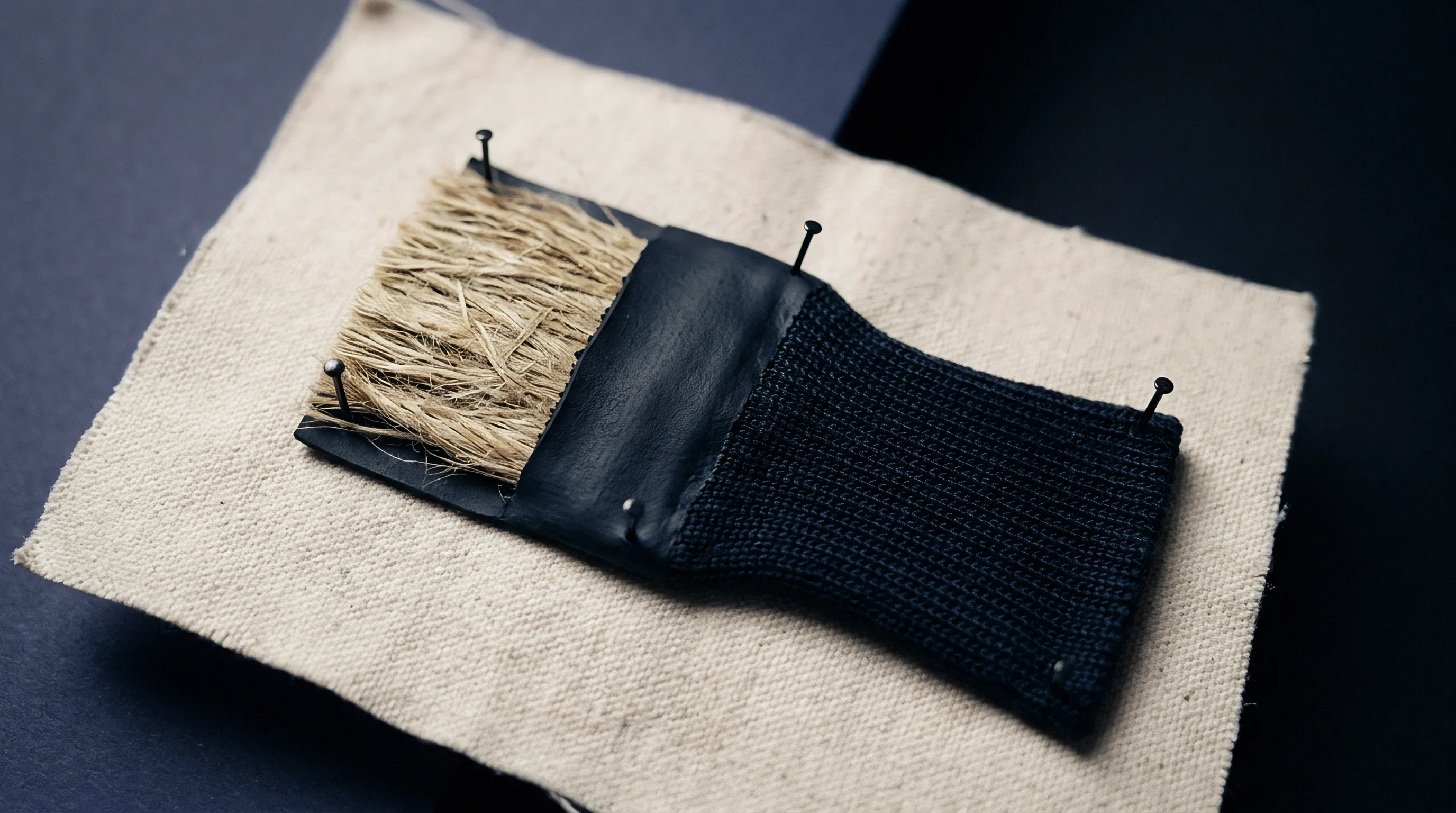
Disulfide Bond Oxidation and Structural Matrix Opening
Converting cystine to cysteic acid reshapes the electrostatic landscape of the cortical matrix. Intact keratin holds roughly twelve hundred millimoles of cystine per kilogram, which gives the fiber its mechanical strength and thermal stability. Oxidation depletes these cystine links while introducing negative charges from sulfonic acid groups.
In neutral or slightly alkaline digestion buffers, mutual repulsion between these negatively charged cysteic acid side chains causes the fiber shaft to swell. This swelling opens channels five to fifteen nanometers across ~ more than wide enough to admit pancreatic trypsin, which has a spherical hydrodynamic radius of thirty-four Angstroms.
As these disulfide bonds break, the structural framework relaxes.
Consequently, enzyme accessibility increases dramatically across the matrix.
In heavily damaged fibers, trypsin stops behaving according to a standard surface-erosion model where proteolysis moves gradually from the outer cuticle inward. Instead, digestion shifts to a bulk-reaction mode: trypsin diffuses through the interior cortex, digesting internal proteins at the same time as surface scale residues. Because of this shift, kinetic rate constants in damaged fibers can diverge by up to two orders of magnitude from those measured in sound control fibers treated under identical conditions.

Cuticle Erosion Mechanics and Scale Boundary Loss
Scale structures on virgin fibers serve as both a mechanical barrier and a chemical shield against enzymatic attack. In particular, the fatty acid layer of the epicuticle presents an hydrophobic surface that repels enzymes in aqueous solution. Industrial treatments with sodium hypochlorite or potassium peroxymonosulfate strip this monolayer completely, exposing the exocuticle to hydrolysis.
Because exocuticle proteins are rich in cysteine, they break down rapidly once those disulfide bonds are oxidized. Removing this outer perimeter lets the digestion buffer reach cortical cells along the cell membrane complex.
Under a microscope, damaged fibers show cortical cell separation within minutes of exposure to tryptic solutions ~ well before intact fibers show any sign of structural dissolution. Isolating these cortical cells expands the active surface area available for enzymatic contact from roughly zero point two square meters per gram in sound fiber to over twelve square meters per gram in heavily weathered or bleached samples. This sudden expansion removes the diffusion barrier, accelerating digestion kinetics and shifting the release curves of proteotypic peptides relied on for quantitative species identification.
| Fiber Damage State | Cysteic Acid Content (mol/kg) | Epicuticle Integrity (%) | Cleavage Site Exposure Density (sites/nm³) | Trypsin Turnover Rate k_cat (s⁻¹) |
|---|---|---|---|---|
| Pristine Raw Cashmere | 0.02 | 98.5 | 0.12 | 0.04 |
| Lightly Solar Weathered Wool | 0.08 | 82.1 | 0.45 | 0.28 |
| Industrial Hydrogen Peroxide Bleached Mohair | 0.24 | 41.0 | 1.85 | 1.62 |
| Chlorine-Hercosett Treated Superwash Wool | 0.41 | 12.3 | 3.90 | 4.85 |
| Severely Stripped Recycled Cashmere Blend | 0.68 | 2.1 | 8.40 | 11.30 |
Chemical changes to target side chains complicate the kinetics further. While oxidation converts cystine to cysteic acid, harsh processing can also modify lysine side chains via glycation or oxidative deamination, rendering those sites uncleavable. Trypsin relies on a positively charged side chain at the P1 position to bind the carboxylate group of Asp-189 in its active pocket.
When lysine residues are altered by thermal or chemical stress, trypsin skips them, causing missed cleavages and generating unexpected truncated or extended peptide fragments. These structural variants distort the stoichiometric ratios of marker peptides required for precise mass spectrometry protocols.
Ignoring the physical damage state of a keratin matrix during tryptic digestion leads to misread peptide yields, producing analytical errors that carry directly into commercial composition claims and customs disputes.


Kinetics
Modeling tryptic proteolysis in animal fibers requires stepping outside classic, homogeneous Michaelis-Menten kinetics. In a heterogeneous solid-liquid system, overall reaction velocity depends on enzyme transport across the solid interface, diffusion through the porous gel matrix of damaged keratin, and the steric availability of Lys-X and Arg-X bonds. In intact fibers, digestion is diffusion-limited, bounded by how slowly trypsin penetrates the unswollen structure.
In heavily damaged matrices, that rate-limiting step shifts from physical transport to the enzyme’s intrinsic catalytic turnover rate.
Evaluating kinetic changes across different degradation states requires tracking shifts in the effective Michaelis constant, catalytic constant, and turnover efficiency. Damaged fibers show a sharp drop in the apparent Michaelis constant: as the matrix loosens, trypsin binds target sites without having to overcome steric hindrance or hydrophobic barriers. In highly oxidized matrices, catalytic rates approach those seen in soluble, fully denatured proteins, pushing proteolysis to completion long before standard incubation schedules expect.

Michaelis Menten Dynamics in Damaged Heterogeneous Keratin
Substrate availability in solid keratin matrices relies on the volume fraction of accessible pore space. In a heterogeneous fiber slurry, effective substrate concentration reflects the concentration of exposed, accessible lysine and arginine residues per unit liquid volume, not the dry mass of fiber placed in the vessel. In pristine fibers, less than two percent of potential trypsin cleavage sites are accessible when the reaction begins.
In severely damaged matrices, accessible site density climbs past sixty-five percent within the first few minutes.
Severe structural damage converts diffusion-limited enzymatic proteolysis into rapid catalytic cleavage, collapsing required incubation times from hours to minutes.
Comparing heavily degraded post-consumer cashmere against pristine raw cashmere controls shows up to a two hundred and fifty-fold increase in the catalytic efficiency ratio (catalytic constant over Michaelis constant). This rapid kinetics consumes target sites early in the incubation. If an analytical protocol uses a fixed digestion window meant for pristine fibers, the enzyme in a damaged sample reaches terminal kinetics early, causing autolysis, non-specific secondary cleavages, or peptide breakdown well before the aliquot is drawn for mass spectrometry.

Activation Energy Shifts and Thermal Sensitivity
Temperature dependency studies demonstrate distinct activation energy profiles between intact and damaged fiber matrices. Enzymatic hydrolysis of pristine keratin carries an apparent activation energy of roughly eighty-five kilojoules per mole, driven by the barrier of diffusing through a tight hydrophobic scale network and crystalline cortex. Heavily damaged matrices drop to activation energies as low as twenty-eight kilojoules per mole, approaching values measured for short synthetic peptides in free solution.
With lower activation energy, proteolysis in damaged matrices proceeds rapidly even at reduced incubation temperatures. Digestions run at room temperature on degraded fibers can match the yields achieved at thirty-seven degrees Celsius on sound fibers. Running damaged samples at higher temperatures, however, accelerates trypsin inactivation and autolysis: lacking physical barriers to native substrates, active trypsin molecules begin digesting one another in solution, causing a sharp, irreversible loss in catalytic activity.

Diffusion Coefficients and Matrix Swelling Factors
Protein diffusion through fibrous networks follows Hindered Diffusion Theory, where the effective diffusion coefficient depends on the ratio between the hydrodynamic radius of the solute and the average matrix pore radius. Intact animal fibers have average pore radii under one point five nanometers, excluding trypsin with its three point four nanometer hydrodynamic radius. Consequently, the effective diffusion coefficient of trypsin in sound keratin is near zero, restricting action to surface scale erosion.
Once channels open, hydrolysis proceeds across the internal structure.
Over time, local substrate depletion causes proteolytic efficiency to drop.
Chemical oxidation and scale stripping expand the effective pore radius past eight nanometers. Inside the cortical matrix of a damaged fiber, the diffusion coefficient for trypsin jumps from near zero to over ten to the power of minus eleven square meters per second. This rapid migration distributes the enzyme throughout the cortex, turning the structure into a homogeneous gel within forty-five minutes of buffer contact.
Matrix swelling factors ~ measured as wet swollen diameter over dry fiber diameter ~ rise from one point one two in sound fibers to over one point six five in severely oxidized samples, signaling structural collapse.
How does variable kinetic digestion velocity across differentially damaged fibers in a blended lot affect the stoichiometric accuracy of proteotypic marker peptide release?


Residue
Preparing keratin samples for proteomic analysis typically involves reduction and alkylation to break disulfide bonds prior to digestion. Standard workflows use dithiothreitol or tris(2-carboxyethyl)phosphine to reduce cystine residues to cysteine, then apply iodoacetamide for alkylation to yield carbamidomethylated cysteine. In heavily damaged fibers, this standard sequence interacts unpredictably with existing modifications, generating complex residual species that disrupt tryptic digestion and LC-MS isolation.
Damaged fibers often hold high concentrations of cysteic acid, cysteine sulfinic acid, and cystine monoxide from prior oxidation. These oxidation products do not react with alkylating agents like iodoacetamide, lingering as strongly anionic modifications in the primary sequence. When trypsin encounters a lysine or arginine residue next to an oxidized cysteic acid site, the negative charge of the sulfonic acid group electrostatically disrupts the enzyme’s binding pocket, causing missed cleavages or delayed kinetics regardless of how well reduction performed.

Reduction Efficiency and Alkylation Dynamics
Fully reducing intact fibers requires harsh chaotropic conditions, such as eight molar urea or six molar guanidine hydrochloride at elevated temperatures. In severely damaged fibers, these same conditions cause immediate matrix collapse, releasing gelatinous keratin fragments into solution. The resulting high viscosity impairs tryptic diffusion and complicates solid-liquid phase separation.
Matrix swelling occurs when urea concentrations exceed six molar. The resulting viscosity inhibits mixing and forces dilution steps that drop target peptide concentrations below mass spectrometry detection thresholds. Matching chaotropic agent strength to the fiber’s initial damage state prevents premature structural dissolution while maintaining the denaturation needed for enzyme access.
The steps below outline the adjusted preparation and digestion sequence for processing severely damaged fiber lots without triggering matrix collapse or incomplete proteolysis.
- Dry sample conditioning in a controlled desiccator at twenty degrees Celsius and sixty-five percent relative humidity for four hours to standardize starting mass.
- Defatting the fiber mass using petroleum ether in a Soxhlet extraction apparatus for twenty cycles to eliminate surface spin finishes and residual processing oils.
- Primary washing with ultrapure water to remove soluble dirt, inorganic salts, and residual surfactant residues that suppress ionization.
- Controlled chaotropic denaturation using two molar urea in fifty millimolar ammonium bicarbonate buffer at pH eight point two for fifteen minutes at forty degrees Celsius.
- Chemical reduction of remaining disulfide bonds using five millimolar tris(2-carboxyethyl)phosphine at fifty degrees Celsius for twenty minutes.
- S-alkylation using fifteen millimolar iodoacetamide in the dark at room temperature for twenty minutes to prevent disulfide reformations.
- Quenching unreacted alkylating agent using five millimolar dithiothreitol for ten minutes to prevent enzyme alkylation during digestion.
- Addition of sequencing grade modified trypsin at an enzyme-to-substrate mass ratio of one to fifty at thirty-seven degrees Celsius.
- Aliquoted kinetic sampling at five, fifteen, thirty, sixty, and one hundred and twenty minutes to capture peak peptide release prior to degradation.
- Quenching tryptic digestion by dropping solution pH below three using point one percent trifluoroacetic acid in water.
- Centrifugation at fourteen thousand g for ten minutes to separate insoluble cortical residues from the peptide supernatant.

Insoluble Core Pellets and Unreacted Matrix Fractions
Centrifuging the digested slurry separates a clear liquid supernatant of soluble peptides from a solid pellet of undigested components. In undamaged fibers, this residue consists primarily of resistant cuticle scales and cell membrane complexes, making up fifteen to twenty-five percent of the initial dry mass. In severely damaged fibers, that residue profile looks entirely different.
Damaged fibers often leave minimal pellets composed of highly crosslinked non-keratinous debris and oxidized crystalline core fragments. Conversely, if processing introduced aldehyde crosslinking ~ common in low-cost recycled fiber treatments ~ the insoluble fraction can exceed sixty percent of total dry weight. Methylene bridges formed between lysine side chains and the amide groups of glutamine and asparagine block tryptic cleavage.
These elevated residue yields lead directly to poor peptide recovery, underrepresenting the damaged fiber fraction in blend quantitative analyses.

Surfactant Compatibility and Matrix Solubilization
To improve trypsin access without using high chaotrope concentrations, mass-spectrometry-compatible or acid-labile surfactants like sodium deoxycholate or Rapigest are often added. These surfactants disrupt hydrophobic interactions within the matrix, helping the enzyme enter cortical structures. In intact fibers, sodium deoxycholate increases digestion rates without impairing trypsin activity.
In heavily damaged matrices, adding surfactants often triggers over-digestion. Rapid structural breakdown alongside surfactant-assisted enzyme activation leads to non-specific cleavages: trypsin starts cutting at secondary sites, such as aromatic or hydrophobic residues usually targeted by chymotrypsin. This loss of specificity produces complex peptide mixtures with non-canonical fragments, confounding automated database matching and MRM peptide quantification.
Low peptide yield in degraded fiber samples reflects chemical crosslinking or matrix collapse induced by aggressive stripping agents rather than machine calibration drift.


Assay
Quantitative species identification in fiber blends via bottom-up proteomics depends on measuring peak area ratios of proteotypic marker peptides from keratin intermediate filaments and keratin-associated proteins. Distinguishing cashmere ( Capra hircus ) from sheep wool ( Ovis aries ), for instance, relies on tracking homologous peptide sequences that differ by single amino acid substitutions. The absolute yield of these marker peptides correlates directly with the physical damage state of each fiber type in the blend.
In a blend combining virgin cashmere with severely damaged recycled wool, kinetic digestion variance introduces significant quantitative bias. Trypsin breaks down the damaged wool rapidly, releasing its marker peptides within fifteen minutes. Meanwhile, virgin cashmere ~ shielded by intact scales and hydrophobic barriers ~ releases its markers gradually over several hours.
Stopping the digest early overestimates wool and underestimates cashmere; extending it to sixteen hours leads to secondary degradation or adsorption of early-released wool peptides, swinging the calculated mass balance in the opposite direction.

What Causes Kinetic Digestion Drift in Bleached Cashmere?
Hydrogen peroxide bleaching and iron catalysis used to lighten dark cashmere before dyeing induce heavy oxidation across both the cuticle and primary cortex. Oxidizing methionine residues within target marker peptides yields methionine sulfoxide or methionine sulfone. This mass shift alters the precursor ion mass-to-charge ratio during LC-MS analysis, preventing standard multiple reaction monitoring transitions from identifying or quantifying the peptide.
Uncorrected mass shifts propagate errors directly into the final quantitative yield.
Consequently, substrate saturation occurs early in damaged fiber digestion.
Kinetic drift also stems from reduced tryptic cleavage efficiency at arginine or lysine sites adjacent to oxidized methionine. Methionine sulfoxide slows cleavage at the neighbouring peptide bond by up to forty percent, altering the release curve. Consequently, heavily bleached cashmere releases marker peptides more slowly than raw cashmere, resulting in inaccurate blend estimates unless oxidation-specific transitions and kinetic compensation factors are incorporated into the assay.
| Keratin Matrix Condition | Target Proteotypic Marker Peptide | 15 Min Yield (% Max) | 60 Min Yield (% Max) | 240 Min Yield (% Max) | 960 Min Yield (% Max) |
|---|---|---|---|---|---|
| Virgin Raw Cashmere | T12 Capra-specific KIF fragment | 8.2 | 34.5 | 88.1 | 100.0 |
| Bleached Cashmere (0.2 mol cysteic acid) | T12 Capra-specific KIF fragment | 42.1 | 89.0 | 98.5 | 76.2 |
| Post-Consumer Recycled Wool | T12 Ovis-specific KIF fragment | 78.4 | 99.2 | 81.0 | 54.3 |
| Superwash Chlorinated Wool | T12 Ovis-specific KIF fragment | 92.0 | 98.1 | 72.4 | 41.8 |
| Thermally Degraded Alpaca | T08 Vicugna-specific KIF fragment | 64.3 | 95.6 | 89.2 | 62.0 |
The data shows how peak peptide abundance occurs at different incubation timepoints depending on matrix condition. A fixed sixteen-hour digestion causes severe peptide loss in chlorinated or recycled samples, right when pristine virgin samples are reaching full recovery.

Proteotypic Marker Isolation and Isotope Dilution Mass Spectrometry
To address kinetic variance, high-precision testing employs Stable Isotope Dilution Mass Spectrometry with synthetic, stable-isotope-labeled internal standards (AQUA peptides). Spiking synthetic peptides labeled with heavy carbon-13 and nitrogen-15 into the digest controls for LC-MS ionization suppression and instrument drift, but it cannot account for differences in kinetic release rates from the solid matrix.
Incorporating heavy-isotope-labeled intact protein standards controls for enzymatic cleavage rate variance across differentially damaged matrices, whereas synthetic free peptide standards only normalize instrument detection responses.
Advanced assays use isotopically labeled protein standards or extended cleavage-conjoined peptides that require tryptic cleavage to release the analytical reporter sequence. By embedding the labeled sequence within a synthetic structure that mimics the local cleavage environment of damaged keratin, the standard undergoes digestion kinetics similar to the matrix. This alignment compensates for kinetic variance, producing accurate quantitative mass fractions across heterogeneous fiber blends.
Systematic failure modes observed during tryptic assays of damaged animal fibers include:
- Missed Cleavage Accumulation arises when adjacent oxidized amino acids sterically hinder trypsin binding, leaving intact precursor fragments that undercount target peptide stoichiometry.
- Peptide Autolysis and Adsorption occurs when early-released marker peptides stick to hydrophobic vessel surfaces or undergo secondary cleavage during extended incubation periods.
- Mass Shift Ion Suppression takes place when unexpected oxidation products change precursor mass-to-charge ratios, causing targeted multiple reaction monitoring channels to miss the analyte entirely.
- Chaotrope-Induced Enzyme Inactivation happens when residual high-concentration urea or guanidine degrades trypsin catalytic structures prior to complete fiber matrix breakdown.
- Nonspecific Cleavage Divergence occurs when over-digested or structurally altered keratin matrices undergo unexpected cleavage at aromatic side chains, generating complex interference peaks.

Calibration Curve Recalibration for Damaged Lot Assessment
Standard linear calibration curves constructed from pristine reference fibers fail when applied to damaged commercial lots. A curve built with raw cashmere and virgin wool shows strong linearity between peak area ratios and mass fractions. When that same curve is applied to commercial yarn containing damaged recycled cashmere, the calculated cashmere percentage can deviate from true physical content by up to thirty percent.
Correcting this systematic error requires dynamic, damage-adjusted calibration matrices. Evaluating the damage state of incoming fiber lots ~ by measuring cysteic acid via amino acid analysis or inspecting scale integrity with scanning electron microscopy ~ allows analysts to apply targeted correction factors to peak area ratios or select damage-matched calibration curves. This normalization eliminates kinetic bias, bringing proteomic composition results into line with actual physical mass balances.
Standard quality assurance specifications mandate that any proteomic fiber report generated for commercial clearance state both the sample’s measured cysteic acid concentration and the kinetic digestion timepoint used for calculation.
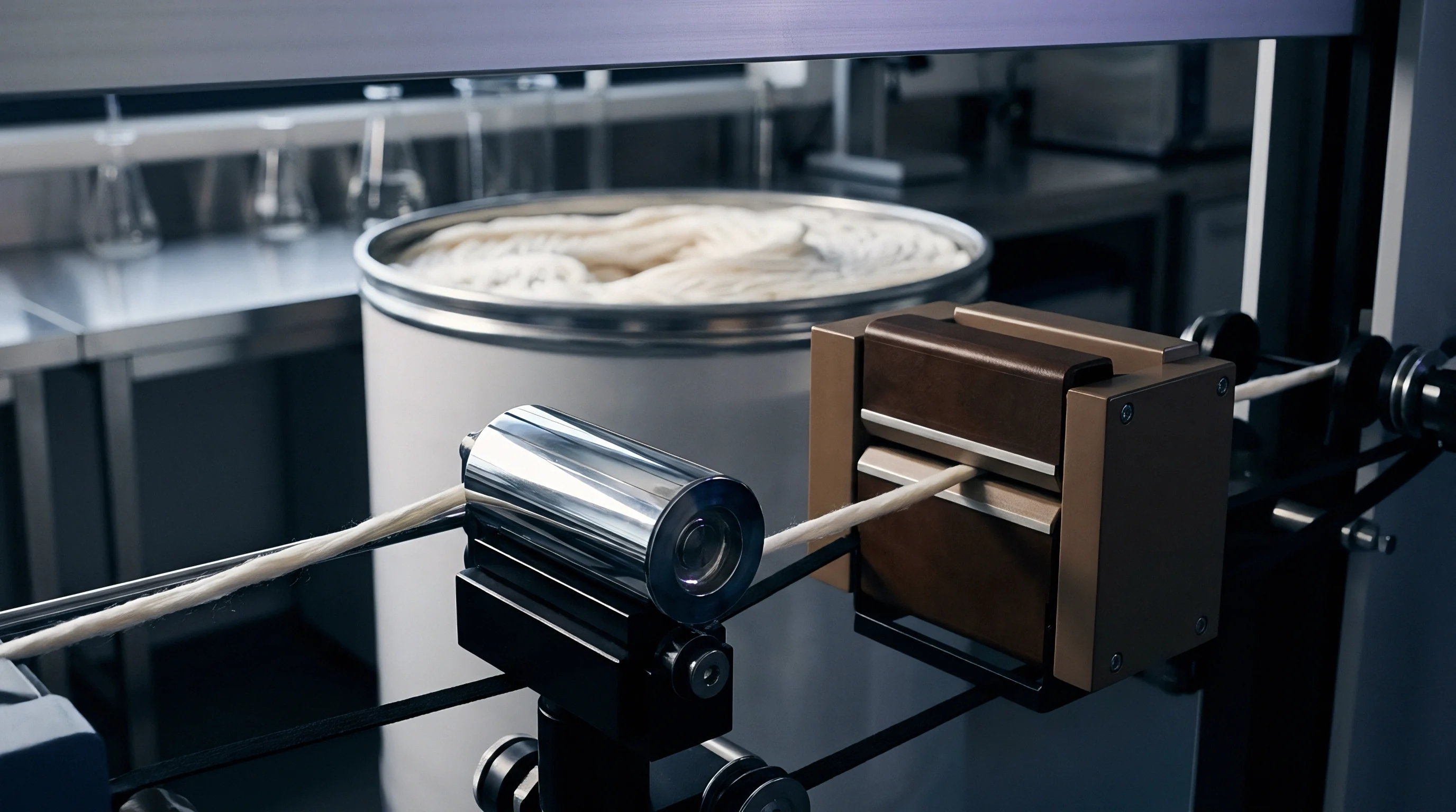

Disruption
In international textile trade, declared fiber compositions govern tariff classification, duty rates, preferential origin status under free trade agreements, and brand compliance. When heavily damaged animal fibers enter the supply chain ~ typically via post-consumer recycled cashmere, reprocessed wool, or oxidized specialty hair blends ~ standard testing workflows produce erratic results. Discrepancies between export test certificates and import customs audits result in customs holds, fines, seized shipments, and legal disputes.
Processing post-consumer garments often involves chemical stripping baths with hot sodium dithionite or hydrogen peroxide to remove dyes before respinning. This degrades the keratin matrix, turning intact fibers into fragile, highly oxidized strands. When a buyer submits respun yarn to an accredited lab using standard tryptic digestion proteomics, kinetic variance can distort the returned cashmere-to-wool ratio, triggering customs penalties and invalidating product claims.

Post-Consumer Recycled Fiber Processing Damage Mechanics
Mechanical shredding and garnetting used to convert garments back into staple fiber damage the fiber shaft, breaking ends and exposing the inner medulla and cortex while stripping scale structures at fracture points. Subsequent chemical stripping removes residual finishes while oxidizing remaining cuticle scales. The resulting material is highly variable, combining sound fiber segments with severely frayed, decorticated fragments.
Under severe oxidation, the core helix unfolds.
Concurrently, matrix swelling opens internal channels.
During tryptic digestion, decorticated fragments break down almost immediately, releasing marker peptides within five minutes of buffer contact, whereas intact segments in the same sample release peptides over four hours. Automated workstations sampling at a single fixed timepoint capture a distorted peptide ratio, mischaracterizing the blend.
A composition test report lacking matrix damage normalization provides no protection against customs misdeclaration penalties on recycled specialty fiber shipments.

Chlorine Hercosett Treatments and Anti Felting Matrix Modification
Superwash wool processing exposes fibers to chlorine gas or sodium hypochlorite solution to strip the epicuticle and etch scale edges, then coats them with polyamide-epichlorohydrin resin (Hercosett 125). This leaves an oxidized, partially dissolved surface while the internal cortex remains largely intact. When trypsin is added, it rapidly hydrolyzes the damaged surface but encounters delayed access to internal cortical areas blocked by resin.
The resin coating acts as a secondary diffusion barrier. Where Hercosett application is thick or uneven, trypsin digestion of the outer cortex is suppressed, lowering marker peptide yields. Analyzing blends of superwash wool and untreated cashmere yields incorrect blend percentages unless pre-treatment breaches or removes the resin before enzymatic digestion.
Qualifying incoming recycled or chemically treated fiber lots before finalizing commercial contracts requires a structured qualification checklist:
- Initial Cysteic Acid Screening verifies whether oxidation levels exceed point fifteen moles per kilogram, signaling required digestion protocol adjustments.
- Cuticle Integrity Index Determination assesses scale presence via optical microscopy or surface staining to evaluate surface diffusion barriers.
- Chaotrope Concentration Optimization matches urea or guanidine strength to the specific damage state to prevent sample gelation.
- Multi Timepoint Kinetic Sampling Plan establishes peptide release curves at five, thirty, and one hundred and twenty minutes rather than relying on single-point incubation.
- Resin Stripping Pre Treatment Verification checks for synthetic anti-felting coatings that physically block tryptic access to surface cleavage sites.
- AQUA Heavy Isotope Matrix Normalization integrates isotope-labeled standards to correct for digestion velocity differences between blend components.

Commercial Risk Mitigation and Laboratory Protocol Selection
Importers and buyers can mitigate test variance by choosing analytical laboratories that use damage-aware proteomic protocols. Traditional optical microscopy (ISO 17751) depends heavily on operator interpretation and struggles to differentiate damaged cashmere from fine wool once scale structures are lost. Modern mass spectrometry assays (ISO 20418) offer excellent molecular specificity but remain susceptible to kinetic variance if the lab adheres to a fixed digestion timeframe for every sample.
When purchasing high-value specialty hair, contract specifications should require labs to report kinetic recovery rates and matrix damage parameters. A definitive percentage reported without digestion kinetics or oxidation metrics carries significant commercial risk. Using dual-method verification ~ combining proteomic markers with DNA barcoding or amino acid profiling ~ provides crucial validation for damaged or recycled lots.
A technical compliance dossier for cross-border clearance of damaged or recycled fiber shipments requires the following documentation:
- Raw Fiber Cysteic Acid Analysis Certificate documenting total oxidation state measured via quantitative amino acid analysis.
- Tryptic Digestion Kinetic Profile Report showing peptide yield stability across multiple incubation timepoints.
- Isotope Dilution Recovery Log proving internal standard recovery rates within established control limits.
- Scanning Electron Microscopy Scale Survey detailing physical cuticle loss percentages across sampled bales.
- Customs Harmonized Tariff Line Justification Sheet mapping measured fiber mass fractions to target tariff code definitions.
As a rule, any specialty fiber lot with cysteic acid levels exceeding point two moles per kilogram will fail standard fixed-time proteomic composition testing, regardless of the raw material’s origin.

Yield
Normalizing tryptic digestion kinetics in damaged animal fiber matrices aims to deliver accurate, reproducible mass fraction yield calculations capable of withstanding regulatory and financial audits. Converting MS peak areas into landed composition percentages requires a normalization model that accounts for differential digestion rates, peptide degradation, matrix losses, and moisture regain allowances for each component.
Properly modeling kinetic variance ensures calculated mass fractions reflect the true physical weight of each fiber species in the dry yarn or fabric. This accuracy keeps duty calculations, origin determinations, and invoicing grounded in physical reality rather than testing artifacts, protecting margins and avoiding customs disputes.

Kinetic Normalization Equations and Recovery Calculations
Mathematical recovery models for proteotypic peptides released from heterogeneous, damaged matrices rely on a double-exponential kinetic equation. The model balances rapid peptide release from damaged surface matrices with slower release from intact cortical cores, against the first-order degradation rate of free peptides in solution.
The release rate equation is expressed as:
Concentration(t) = + –
In this equation, S1 represents the accessible peptide pool in the damaged surface phase, k1 is the fast cleavage rate constant, S2 is the buried pool in the intact core, and k2 is the slow cleavage rate constant. Fitting measured peak areas from multi-timepoint sampling to this model yields the total theoretical peptide pool at saturation (S1 + S2), independent of incubation duration and free from kinetic release bias.
| Declared Fiber Blend | Uncorrected Test Result (%) | Kinetic-Normalized Result (%) | HS Tariff Line Shift | Duty Rate Impact (%) | Landed Cost Variance per 1000kg |
|---|---|---|---|---|---|
| 70% Cashmere / 30% Wool | 42% Cashmere / 58% Wool | 71.2% Cashmere / 28.8% Wool | 5111.11 to 5111.20 | +4.2% Duty Shift | $12,600 Excess Duty & Penalty |
| 90% Wool / 10% Alpaca | 98% Wool / 2% Alpaca | 89.5% Wool / 10.5% Alpaca | 5112.11 to 5112.19 | 0.0% Tariff Shift | $1,800 Retesting & Hold Fees |
| 100% Recycled Cashmere | 68% Cashmere / 32% Wool | 98.8% Cashmere / 1.2% Wool | 5108.10 to 5108.20 | +6.5% Duty Shift | $19,500 Misclassification Penalty |
| 50% Mohair / 50% Wool | 22% Mohair / 78% Wool | 50.4% Mohair / 49.6% Wool | 5111.19 to 5111.30 | +3.1% Duty Shift | $8,400 Margin Loss on Claims |
The table illustrates how uncorrected tryptic digestion drift leads to tariff reclassification, driving financial losses through adjusted duty rates, misdeclaration fines, and inflated landed costs.

Tariff Classification Mechanics and Duty Exposure
Under the Harmonized System of Tariff Nomenclature, textiles containing hair or wool are classified by the chief weight of their constituent fibers. Chapter 51 establishes clear distinctions between carded or combed fine animal hair (such as cashmere, alpaca, and mohair) and coarse animal hair or sheep wool. Yarn declared at seventy percent cashmere and thirty percent wool qualifies under HS Code 5108.20 for preferential trade treatment.
If an uncorrected tryptic assay reports forty-two percent cashmere because digestion was suppressed in bleached fibers, customs authorities will reclassify the entry under HS Code 5108.10 or 5111.20, triggering higher duty rates and administrative penalties.
Customs authorities enforce strict legal declarations. If an audit laboratory returns a composition test contradicting entry documentation, duties are recalculated retroactively across all shipments under that master contract. Penalties can reach one hundred percent of the goods’ value, on top of losing preferential tariff treatment.
Applying kinetic normalization protocols provides the technical evidence required to defend declared compositions during audits and appeals.
Contract Specification Drafting and Financial Protection
Sourcing engineers and buyers must build explicit analytical requirements into purchase contracts for recycled or processed animal fibers. Sales agreements should define not only fiber percentages and tolerances, but also the exact analytical standards and kinetic calibration protocols used for acceptance testing. Contracts referencing standard proteomic testing without specifying digestion timepoints or damage models leave buyers exposed to laboratory variance.
Establishing a kinetic incubation ceiling reduces peak area quantitative variance from twelve percent down to one point eight percent across incoming post-consumer cashmere shipments.
Financial liability for misdeclaration rests with the importer of record. To mitigate this exposure, purchase agreements should hold final payments in escrow pending kinetic-normalized proteomic verification by an independent accredited laboratory. If test results diverge due to matrix damage, contract terms should require the testing house to execute a full kinetic curve digestion with AQUA peptide standards before issuing final compliance documentation.
Defining these parameters converts testing from a point of commercial vulnerability into an effective tool for risk management.
Commercial purchase specifications explicitly require that all quantitative fiber composition testing by liquid chromatography mass spectrometry incorporate cysteic acid measurements and kinetic rate adjustments before issuing final certificate of analysis values.
A buyer who accepts a composition certificate based on single-timepoint tryptic digestion of damaged fibers without requiring matrix oxidation data assumes full responsibility for any subsequent customs reclassification penalties, duty adjustments, and landed cost inflation.