Standardizing Trypsin Digestion Parameters for Cashmere Identification
Standardized trypsin digestion at pH 8.2 and thirty-seven degrees for sixteen hours yields diagnostic peptides separating cashmere from wool by liquid chromatography.

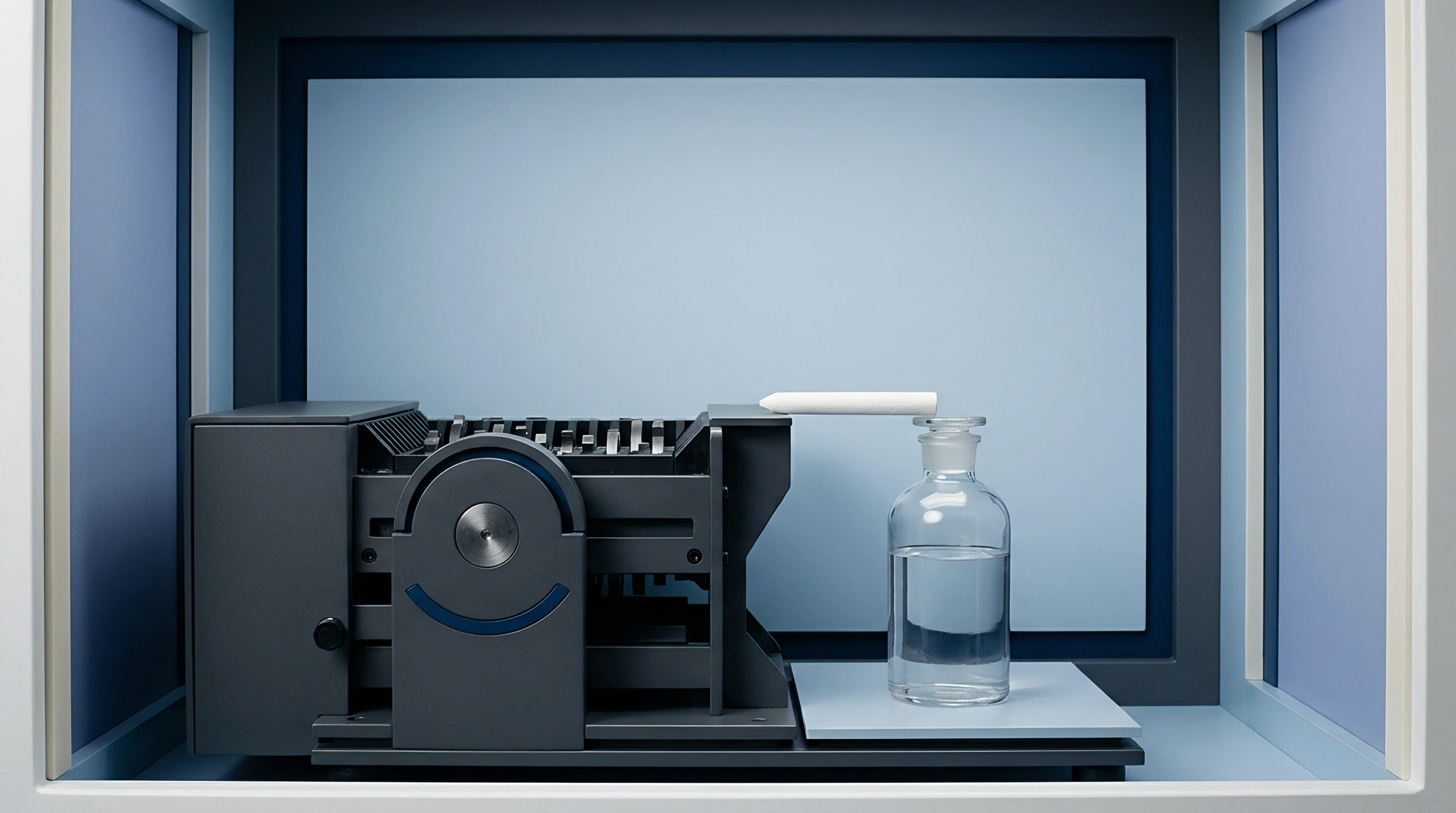
Bath
Proteomic mass spectrometry requires strict boundary conditions during sample solubilization to release diagnostic keratin chains. Raw cashmere guard hair and down fibres arrive coated in suint, wax, and processing lubricants that inhibit proteolytic activity. Technicians wash scoured samples three times in petroleum ether, rinse with deionized water, and dry the fibre mass at forty degrees Celsius to constant weight.
Without thorough degreasing, solvent-insoluble residues blind analytical columns. Solubilizing the resilient disulfide matrix demands a concentrated denaturing environment containing eight molar urea, fifty millimolar ammonium bicarbonate, and ten millimolar dithiothreitol.
Dithiothreitol cleaves the intra-chain and inter-chain cystine linkages across intermediate filament proteins. Incubation proceeds at fifty-six degrees Celsius for forty-five minutes. Temperature excursions past sixty degrees cause carbamylation of lysine and arginine residues, altering the target cleavage sites.
Once disulfides reduce to free sulfhydryls, alkylation prevents bond reformation. Iodoacetamide serves as the standard capping agent at a concentration of fifty-five millimolar. Technicians hold the solution in darkness at room temperature for forty minutes.
Light exposure induces iodine radical reactions with tyrosine residues, skewing subsequent mass readings.
Trypsin hydrolyzes ninety-eight percent of accessible peptide bonds when maintained at pH 8.2 and thirty-seven degrees Celsius for sixteen hours.
Diluting the urea concentration below one molar protects enzyme vitality before adding sequencing-grade modified trypsin. High urea concentrations denature the enzyme, halting proteolysis prematurely. Ammonium bicarbonate buffer at fifty millimolar maintains the digestion liquor at pH 8.2.
Technicians meter sequencing-grade trypsin at an enzyme-to-substrate mass ratio of one to fifty. Pure cashmere lots digest predictably under these conditions. The digestion vessel remains in an orbital shaking incubator at thirty-seven degrees Celsius for sixteen hours.
Terminating the reaction requires immediate acidification. Technicians introduce neat trifluoroacetic acid to bring the total solution volume to one percent acid, lowering the bath to pH 2.0. Proteolysis halts instantly.
Centrifugation at fourteen thousand gravities for ten minutes pellets undigested cortical remnants.

Stepwise Preparation Sequence
- Solvent degreasing removes spinning oils and residual waxes through three successive washes in petroleum ether followed by desiccator drying.
- Disulfide reduction breaks cross-linked cystine bridges in eight molar urea buffered with fifty millimolar ammonium bicarbonate at fifty-six degrees.
- Thiol alkylation caps liberated sulfhydryl groups using fifty-five millimolar iodoacetamide during a forty-minute dark incubation.
- Buffer dilution lowers the denaturant below one molar urea so the added endopeptidase retains enzymatic activity.
- Acid quenching halts proteolysis after sixteen hours by driving the pH down to two with trifluoroacetic acid.
| Reagent Stage | Chemical Component | Working Concentration | Thermal Setting | Holding Duration |
|---|---|---|---|---|
| Degreasing | Petroleum ether | 100 percent | 22 degrees C | 30 minutes |
| Solubilization | Urea | 8.0 mol/L | 22 degrees C | 15 minutes |
| Reduction | Dithiothreitol | 10.0 mmol/L | 56 degrees C | 45 minutes |
| Alkylation | Iodoacetamide | 55.0 mmol/L | 22 degrees C (dark) | 40 minutes |
| Proteolysis | Sequencing-grade trypsin | 1:50 mass ratio | 37 degrees C | 16 hours |
| Quenching | Trifluoroacetic acid | 1.0 percent v/v | 22 degrees C | Instant |
A vessel left unheated yields uncleaved protein sheets that choke microfluidic injection loops.
Strand
Keratins divide into acidic Type I and basic-to-neutral Type II intermediate filament assemblies. The cortex of Capra hircus laniger and Ovis aries contains near-identical macromolecular architecture, yet single-point amino acid substitutions differentiate the species. Trypsin acts exclusively at the carboxyl side of lysine and arginine residues, except when followed by a proline.
Complete digestion yields hundreds of short peptides, but only a handful contain species-discriminating polymorphisms. In Type I microfibrillar keratin K33A, cashmere fibres produce a diagnostic fragment designated as T8, containing a threonine residue. Sheep wool substitutes a serine residue at the equivalent position, displacing the observed mass-to-charge value.
Scanning electron microscopy historically separated these fibres by scale frequency and scale height. Wool exhibits scale heights exceeding 0.55 micrometres with prominent coronal patterns. Dehaired Chinese and Mongolian cashmere down exhibits scale heights below 0.40 micrometres with cylindrical margins.
Mechanical stretching, descaling enzymes, and chemical smoothing wash away these surface differences, causing optical microscopy to misclassify sheared merino wool as pure down. Mass spectrometry bypasses the cuticle entirely by reading the primary amino acid sequence of the cortical proteins.
Capra hircus alpha-keratin contains a leucine residue where Ovis aries substitutes isoleucine along the identical sequence segment.
Enzymatic efficiency determines whether species-specific marker peptides appear in quantifiable abundance. Under-digested samples retain high-molecular-weight fragments that elute across wide retention windows, obscuring low-abundance biomarkers. Over-digested samples or prolonged incubations provoke non-specific cleavages along chymotryptic-like sites, destroying target peptides.
Yak down introduces a third diagnostic profile, derived from Bos grunniens keratin K36. Yak hair yields distinct acidic fragments that allow three-way separation in complex mixtures.
Digestion Failure Modes
- Enzyme autolysis generates extraneous tryptic fragments that crowd mass spectra when incubation temperatures exceed thirty-eight degrees Celsius.
- Incomplete reduction leaves disulfide bonds intact, blocking enzyme access to internal cleavage sites within the microfibrillar matrix.
- Excessive alkylation causes off-target modification of methionine, histidine, and lysine amines, shifting anticipated mass-to-charge ratios.
- Secondary chymotryptic activity clips peptides at tyrosine and phenylalanine bonds after prolonged digestion windows past twenty-four hours.
Analytical consistency rests on reproducing identical digestion kinetics from lot to lot across varying raw hair origins. Cashmere sourced from the Alashan plateau presents a tighter cortical packing than down from outer Mongolia, demanding rigid adherence to the sixteen-hour digestion window to liberate equivalent peptide concentrations.


Peak
Triple quadrupole mass spectrometers resolve diagnostic ions through selected reaction monitoring. The method pairs reversed-phase liquid chromatography using a C18 column with electrospray ionization. Mobile phase A consists of 0.1 percent formic acid in HPLC-grade water.
Mobile phase B contains 0.1 percent formic acid in acetonitrile. A shallow linear gradient spanning five to thirty-five percent organic solvent over twenty-two minutes separates the tryptic digest efficiently. The diagnostic cashmere peptide yields a doubly charged precursor ion at mass-to-charge 847.4, while the sheep wool homologous peptide appears at 840.4.

Can Peak Area Ratios Separate Admixtures below Five Percent?
Quantifying intimate wool mixtures in declared pure cashmere down relies on calibration curves generated from synthetic peptide internal standards. Heavy isotope-labeled standard peptides spiked into the digest compensate for ion suppression during electrospray ionization. The limit of detection for sheep wool marker peptides stands at 0.5 percent by total protein weight.
The limit of quantification reaches 1.0 percent. When sheep wool content drops below three percent, ion counts near the detection threshold, requiring longer signal integration times.
| Fibre Origin | Keratin Subunit | Peptide Sequence | Precursor Ion (m/z) | Product Ion (m/z) |
|---|---|---|---|---|
| Capra hircus (Cashmere) | K33A Type I | TGVDVQLQISLTR | 716.40 2+ | 847.48 y8+ |
| Ovis aries (Wool) | K33A Type I | SGVDVQLQISLTR | 709.39 2+ | 847.48 y8+ |
| Bos grunniens (Yak) | K36 Type I | TGVDVQLQVGLTR | 701.39 2+ | 817.47 y8+ |
| Capra hircus (Cashmere) | K31 Type I | LCEACFFDK | 573.74 2+ | 741.34 y5+ |
| Ovis aries (Wool) | K31 Type I | LCEACLFDK | 580.75 2+ | 755.36 y5+ |
Synthetic internal standards correct for recovery variations across extraction batches. Uncalibrated peak area ratios drift up to twelve percent between individual chromatographic runs when source temperature varies by five degrees. A laboratory running raw area integration without internal normalization produces conflicting results on identical yarn packages.
Uncorrected baseline drift in low-tier spectrometers leads to false identification of wool markers in authentic down, triggering unjustified shipment rejections and lost deposits.


Bleach
Industrial mills treat dehaired lots with hydrogen peroxide to lighten pigmented hair before yarn spinning. Strong oxidative processing damages cystine residues, converting disulfides into cysteic acid. Cysteic acid groups introduce strong negative charges along the keratin backbone that alter protein folding.
Oxidized keratins resist enzymatic cleavage, slowing trypsin kinetics. The resulting mass chromatograms show attenuated marker peaks and elevated background noise.
Will Industrial Bleaching Suppress Diagnostic Ion Signals?
Severe bleaching oxidizes methionine residues to methionine sulfoxide, adding sixteen mass units to target peptides. Analytical screening configurations that monitor only unmodified mass transitions miss these altered fragments entirely. The calculated cashmere content then drops artificially.
Laboratories evaluating bleached or stripped lots must monitor both native and oxidized transitions for each peptide marker. A yarn damaged by over-bleaching requires twenty-four hours of digestion rather than sixteen to release equivalent peptide mass.
Severe peroxide treatment damages keratin chains so thoroughly that tryptic cleavage produces unidentifiable sub-fragments.
Reductive bleaching using sodium dithionite causes less oxidative side-chain damage, preserving predictable cleavage sites. Acid chlorination, applied during shrink-resist treatments, degrades the cuticle while partially cleaving cortical peptide bonds before the sample ever reaches the testing bench. The analyst checks the physical condition of the lot before digestion starts.
Pre-Digestion Assessment Protocol
- Alkaline solubility testing screens for severe oxidative degradation by measuring fibre dissolution in 0.1 molar sodium hydroxide at sixty-five degrees.
- Cysteic acid quantification via Fourier transform infrared spectroscopy tracks sulfoxidation levels across the 1040 reciprocal centimetre band.
- Urea bisulfite solubility determines cortical protein cross-linking density prior to setting the enzyme-to-substrate ratio.
- Visible spectrum reflectance confirms chemical de-pigmentation on white cashmere lots that originated from dark grey raw stock.
Spinning mills frequently claim that chemical treatments dissolve the wool markers while leaving cashmere markers unharmed.
Margin
Cross-border customs entries face steep tariff reclassifications when lab results contradict paperwork declarations. Cashmere fibre sits under Harmonized System heading 5102.11 with an import tariff duty rate around four percent in major destination markets. Carded or combed sheep wool under heading 5105.29 carries different tariff lines and quota restrictions.
Intimate wool admixtures disguised as pure cashmere alter duty computations and risk trade fraud actions.
Raw white cashmere down trades at eighty to one hundred dollars per kilogram, while fine merino wool of matching seventeen-micron diameter trades at fifteen dollars per kilogram. An admixture carrying fifteen percent merino wool saves the yarn spinner over nine dollars per kilogram in raw material outlays. On a forty-thousand-meter spinning run of gauge twelve knitwear, an undeclared ten percent wool component diverts thousands of dollars in input expenses directly to processor profits.
Proteomic testing costs three hundred to five hundred dollars per lot, a trivial expense compared to the financial exposure of an impounded commercial container.
Under the Wool Products Labeling Act, undeclared sheep fibre exceeding three percent by mass subjects the entire lot to immediate customs seizure.
The standard tolerance for accidental contamination in textile labeling laws stands at three percent. When LC-MS/MS confirms deliberate inclusion of seven percent merino wool in goods marked one hundred percent cashmere, civil penalties accompany immediate product destruction. Sourcing contracts that specify ISO 1833 chemical dissolution fail to protect the buyer because both fibres dissolve identically in alkaline hypochlorite.
The buyer specifies proteomic digestion parameters in purchase specifications.
Purchase contracts stating that fibre identification must follow proteomic mass spectrometry with quantification tolerances under two percent bind the spinner to traceable laboratory audits.